
The Four Views
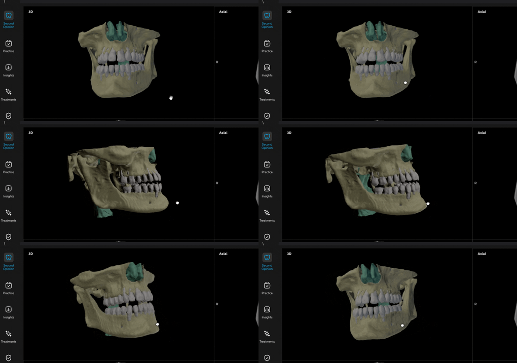
- 3D — a full, rotatable model of the jaw and skull.
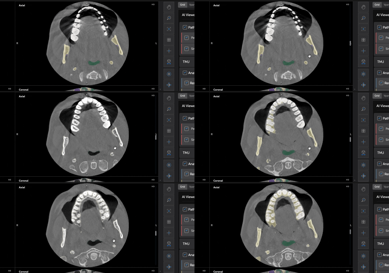
- Axial — a horizontal slice, as if looking down from above.
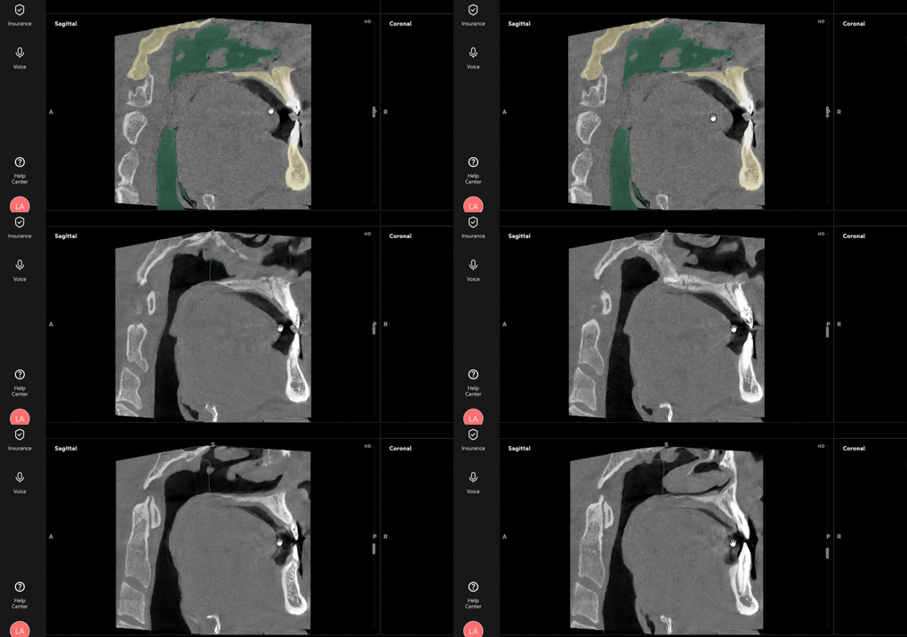
- Sagittal — a vertical slice from the side.
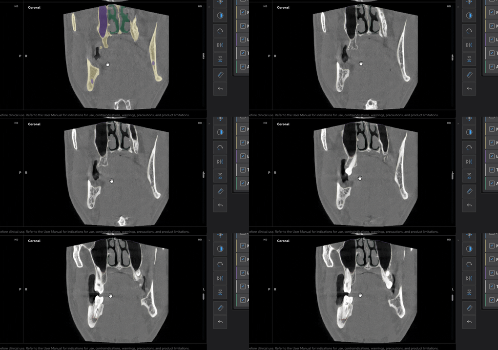
- Coronal — a vertical slice from the front.
Navigating the Grid
- Scroll with your mouse wheel to move through Axial, Sagittal, or Coronal slices.
- Double-click any panel to enlarge it to full screen. Double-click again to return to the four-panel Grid.
- Click and drag on the 3D panel to rotate the model freely.
Color-Coded Anatomy
|
Color |
Structure |
|
Red |
Pathology (AI-detected findings) |
|
Blue |
Restorations (root canals, implants, bridge pontics) |
|
Yellow |
Maxilla and mandible |
|
Purple |
Nerve canals + Sinus |
|
White |
Teeth |
|
Green |
Airway |
Best Practices
- Use the color coding as a quick visual anchor when a scan first loads, before diving into specific findings.
- Enlarge a single panel (double-click) when you want to study one view closely without the others as visual noise.