
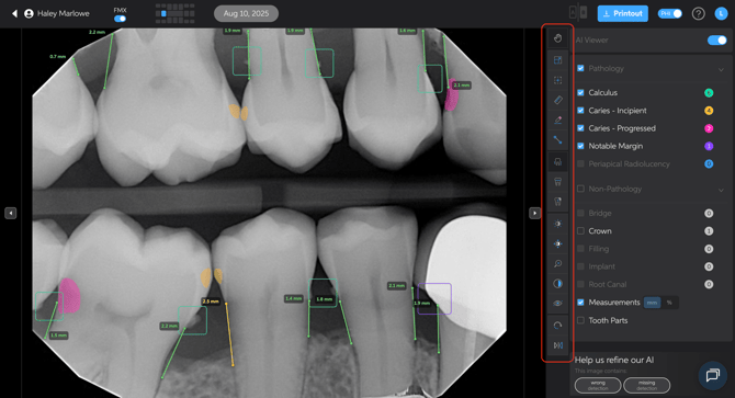
Using the toolbar
The Pearl toolbar offers many useful tools for viewing, manipulating and adjusting images for various use cases.
Jump to Feature
- Full Screen
- Reset View
- Ruler
- Edit
- Edit Measurements
- Measurements
- Tooth Parts
- Affected
- Brightness, Contrast, Zoom
- Invert
- Image Enhance
- Rotate
- Flip
Full Screen
- Use this to enlarge the image for better review and case presentations.
Reset View
- If you’ve moved the image while reviewing a specific area, this button will reset the view back to the original position.
Ruler
- Measures endpoints to determine working lengths for endo, pathologies, implant space placements
Edit
- While the AI uses computer vision to highlight areas of interest, you remain in control of the diagnosis and treatment decisions.
- If you disagree with an AI detection (false positive or false negative), you can modify it:
- Remove a detection
- Restore a removed detection
- Manually add a detection
Click Here for more information on the Edit tool
Edit Measurements
- Allows you to adjust measurements related to bone levels for a more precise analysis.
- Enable edit measurements, then click + drag measurement endpoints to desired location.
Measurements
- Enables or disables bone measurement overlays on the image.
- This can also be done by checking or unchecking on the control panel further to the right
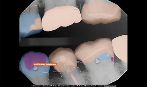
Tooth Parts
- A case presentation tool that helps educate patients by converting grayscale X-rays into a color-coded view:
- White = Enamel
- Green = Dentin
- Purple = Nerve/pulp
- Blue = Cementum
- This visual aid helps patients understand the severity of decay or other conditions and why treatment is needed.
- Can be useful in explaining “watch” areas vs. areas requiring immediate treatment.
Affected
When enabled, the feature calculates the percentage of the healthy crown affected by:
- Existing restorations
- Caries (decay)
- Missing tooth structure
These areas are combined to produce a single percentage value, commonly referred to as Decayed, Missing, Filled (DMF).
Note: Dental crowns covering an entire tooth will not yield an affected percentage because the entire tooth structure is considered altered.
Click Here for more information on the Affected feature
Brightness, Contrast, and Zoom
- Brightness & Contrast: Adjust for better visualization of specific areas.
- Zoom: Use the slider or the scroll wheel on your mouse to zoom in and out.
- Reset View: Returns image settings to default.
Invert
- Reverses the grayscale image, which can sometimes enhance visibility of certain conditions.
Image Enhance
- Adjusts the image clarity/sharpness:
- Lower clarity/lower number = Softer appearance
- Higher clarity/higher number = More defined and crisp image
- Set the enhancement level once per device, and it will stay for all images moving forward.
- Different providers can customize their preferred settings per device using toolbar
- Global setting can be applied in “Customize interface”
Rotate
- Rotate clockwise or counterclockwise and confirm position to reprocess
Flip
- Flip horizontally or vertically and confirm to reprocess